
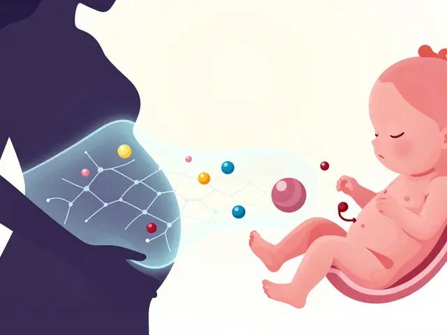
The idea that the Placenta is a perfect shield protecting the fetus from everything outside is a myth we need to discard quickly. In reality, this organ acts more like a selective filter than a wall. We know this because history taught us a harsh lesson. In the late 1950s and early 1960s, the drug Thalidomide, intended to calm morning sickness, caused severe birth defects globally. This tragedy proved that pharmaceutical compounds can breach the barrier between mother and baby.
Understanding the Barrier: It Is Not Static
You might assume the barrier is always the same thickness and strength throughout pregnancy. That assumption is dangerous. By the time you reach full term, the Placenta weighs about 500 grams and covers roughly 15 square meters. However, its permeability changes constantly. During the first trimester, when the organs are forming, the tissue is far more porous than in later stages. This means a medication taken at eight weeks has a different chance of reaching the developing system than one taken at thirty-eight weeks. The barrier evolves alongside the pregnancy itself.
Why does this variation matter? Think about the timing of development. If a critical window closes before a transporter kicks in, damage can occur silently. Experts like Dr. Susan Fisher have noted that the placenta is an adaptive organ responding to maternal conditions. Ignoring this dynamic nature leads to underestimating fetal exposure.
Mechanisms of Transfer: How Drugs Move
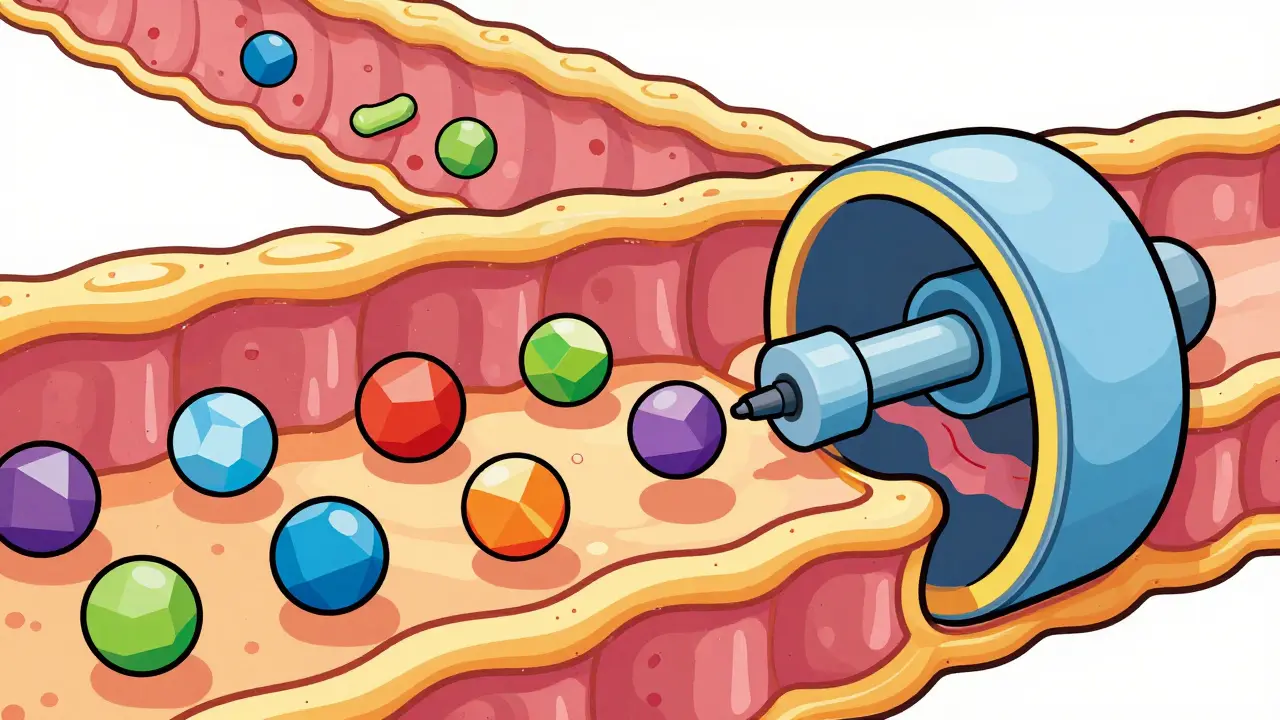
So, how exactly does a molecule get from your blood to the umbilical cord? There are distinct pathways involved here. Most medications rely on passive diffusion. Imagine dropping sugar into tea; eventually, it spreads out naturally until concentrations equalize. Small, lipid-soluble molecules follow this rule easily. Substances like ethanol pass through rapidly, often achieving the same concentration in fetal blood as in maternal blood within an hour.
Not everything moves so freely, though. Larger or water-loving molecules face resistance. Insulin, a large protein, barely crosses at all. Instead of relying on luck, some drugs hijack biological systems called transporters. These act like pumps or gates embedded in the cell walls. One crucial player is P-glycoprotein. This protein actively pushes certain drugs back into the maternal circulation before they reach the fetus. It works like a security guard ejecting unwanted visitors.
Factors Determining Exposure Levels
If you want to predict whether a drug affects a fetus, you cannot look at the chemical structure alone. Several variables dictate the final outcome. First, consider molecular size. Generally, anything smaller than 500 Daltons has a much easier time crossing than larger complexes. This small threshold increases transfer likelihood by about 70%.
Lipid solubility plays another massive role. Compounds that dissolve well in fats travel through cell membranes faster. If a substance has a high log P value, meaning it loves fat over water, expect higher fetal levels. Protein binding adds complexity too. Only the free fraction of a drug moves across the membrane. Warfarin binds tightly to proteins, leaving very little unbound material available to cross, despite having favorable chemical properties otherwise.
Gestational age shifts the equation again. Early on, tight junctions between cells are not fully developed. You might see two to three times more permeability in the first trimester compared to term. As the pregnancy progresses, efflux transporters like P-glycoprotein mature and become more active, often reducing the amount of drug that successfully transfers.
Drug Classes and Their Specific Risks
Different categories of medicines behave differently in this environment. Psychiatric medications often cross quite readily. Selective serotonin reuptake inhibitors, such as sertraline, have shown cord-to-maternal ratios near 1.0. This means almost equal amounts exist in both parties. For some babies, this exposure correlates with transient neonatal adaptation syndrome occurring in about 30 percent of cases shortly after birth.
Pain management presents another challenge. Opioid medications like methadone achieve fetal concentrations reaching 65 to 75 percent of maternal levels. Because the fetus metabolizes these substances slowly, withdrawing from them after birth can trigger Neonatal Abstinence Syndrome. Statistics suggest this occurs in up to 80 percent of exposed infants born to mothers on maintenance therapy.
Antiepileptic drugs show a wider range of behaviors. Phenobarbital moves easily, creating nearly identical concentrations in both circulations. Valproic acid also crosses with high efficiency. However, this convenience comes with risks. Major congenital malformation rates associated with valproic acid sit around 10 to 11 percent, significantly higher than the baseline rate seen in the general population.
HIV treatments offer a fascinating counter-example. Many antiretroviral drugs interact heavily with the P-glycoprotein pump. Lopinavir, for instance, reaches only 60 percent of maternal concentration in the fetus compared to zidovudine which hits 95 percent. This difference proves that transporter activity can drastically limit fetal exposure even among effective antiviral therapies.
Modern Research and Future Tools
We cannot test every hypothesis on human pregnancies. To understand these mechanisms better, scientists use advanced models. Dually perfused human placenta models allow researchers to mimic blood flow and measure transfer rates in real-time. More recently, Placenta-on-a-Chip technology has emerged. These microengineered systems replicate the microscopic architecture of the organ. Studies using this tech show glyburide transfer rates matching ex vivo results closely, validating their accuracy for prediction.
Nanotechnology is also entering the conversation. Some researchers hope to design drug carriers that specifically target the placenta without entering fetal tissue indiscriminately. While promising, concerns remain about nanoparticles accumulating in placental tissue and causing inflammation. The industry invests significantly here, aiming for safer delivery methods that minimize unintended exposure.
Despite these advances, species differences create translation hurdles. Mouse placentas are structurally different from humans. They show greater permeability to certain agents, sometimes misguiding toxicity predictions if relied upon exclusively. Human data remains king when making clinical decisions.
Clinical Management and Guidelines
For clinicians managing pregnant patients, monitoring becomes essential when narrow therapeutic index drugs are involved. Therapeutic drug monitoring ensures blood levels stay within safe ranges, preventing both under-treatment of the mother and overdose of the fetus. The American College of Obstetricians and Gynecologists recommends this approach for medications where small dosage changes significantly alter outcomes.
Regulatory bodies have evolved their requirements as well. The FDA now mandates specific placental transfer data for new applications targeting women of childbearing potential. Guidance documents from 2018 specify quantitative metrics needed for developmental toxicity studies. This shift reflects a recognition that assuming safety without data is no longer acceptable practice.
Patients deserve transparency during decision-making. Discussing the specific transport properties of prescribed Medications allows for informed consent. Understanding that a barrier exists but is imperfect helps set realistic expectations regarding treatment benefits versus potential risks to development.

Owen Barnes
April 1, 2026 AT 07:39The historical context of thalidomide remains paramount in understanding modern obstetric pharmacology.
We often forget how quickly the scientific community adapted after that tragedy occured globally.
The tissue structure changes significantly during gestation which alters permeability rates constantly.
It is crucial to acknowledge that the organ functions as a selective filter rather than an impenetrable wall.
Many clinicians still operate under the misconception that maternal medication does not impact fetal development.
Early trimester exposure carries substantially higher risks due to the porous nature of developing junctions.
Protein binding characteristics dictate how much free drug is available to cross into the umbilical circulation.
Lipophilicity plays a major role in determining which molecules can traverse cell membranes efficiently.
Smaller compounds typically encounter less resistance compared to larger macromolecules attempting transfer.
Active transport mechanisms like p-glycoprotein serve as biological pumps ejecting unwanted substances back to mother.
Understanding these pathways helps explain why certain antiretrovirals succeed where others fail in preventing vertical transmision.
We must consider molecular weight thresholds when predicting potential teratogenic outcomes during pregnancy management.
Clinical guidelines have shifted toward requiring quantitative metrics for new drug applications involving childbearing women.
Transparency regarding specific transport properties allows patients to make fully informed decisions about their care options.
Ignoring the dynamic nature of the barrier leads to dangerous underestimations of fetal exposure levels today.
Future research using placenta on a chip models promises to refine our predictive capabilities significantly soon.
Molly O'Donnell
April 2, 2026 AT 04:58Ignoring the science here could lead to another global health catastrophe repeating history.
Rocky Pabillore
April 4, 2026 AT 03:44Some individuals seem to lack the foundational knowledge required to discuss such nuanced physiological processes correctly.
It appears the nuances of efflux transporters are often overlooked by those who prefer sensationalism over evidence.
Rod Farren
April 4, 2026 AT 23:40Regarding the lipid solubility parameters, the log P value is critical for assessing transplacental passage rates.
Efflux mechanisms mediated by ABCB1 transporters effectively reduce intracellular accumulation in the syncytiotrophoblast layer.
Bioavailability profiles differ significantly depending on the specific gestational window involved.
Pharmacokinetic modeling requires precise input regarding molecular weight and ionization states.
James DeZego
April 5, 2026 AT 03:41Great breakdown of the ABCB1 mechanisms you mentioned there :)
It really highlights why P-gp activity is so vital for protection.
Julian Soro
April 5, 2026 AT 17:27Totally agree with the emphasis on protective transporters keeping harmful agents at bay.
It gives me hope for better drug design in the near future.
Jenny Gardner
April 7, 2026 AT 12:08This information is absolutely fascinating!!
The concept of the barrier evolving throughout pregnancy changes everything we thought we knew!!
We must demand stricter regulations!!
Sharon Munger
April 9, 2026 AT 10:44i think strict regulation is key.
we need better data before approval
its a big step forward though
glad to see the research moving forward
Cara Duncan
April 10, 2026 AT 04:46Wow thank you for the detailed history lesson 😮👏
The part about the porous junctions really opened my eyes 👁️💡
Safety comes first always 🛡️❤️
Cullen Zelenka
April 10, 2026 AT 23:44Progress in microengineering models offers a promising outlook for safer therapeutic options.
Continued investment in human data collection ensures clinical decisions remain evidence based.